Words
Published
The term Retinoschisis cover a series of disorders of the eye in which the retinal layer at the back of the eye splits into two separate layers. One of these disorders – juvenile retinoschisis – is so called because it appears around the age of ten or so years.
The term juvenile is also used to differentiate the retinoschisis from another called “senile retinoschisis” which only affects those in their forties. The two types of retinoschisis, although having the same property of splitting of the retinal layer, have different causes and outcomes.
The main area where the retinal splitting occurs is in the most sensitive part of the retina capable of discerning minute detail and importantly colour – the macula.
If the two layers increasingly stray apart then the space between them can fill with blood. The blood can then find its way into the vitreous gel in the centre of the eye and any material between the lens and the retina will lead to cobweb like images or “floaters” being imagined because of blockage of the light.
It is possible that splitting of the layers may lead to the very serious condition of retinal detachment, but this is relatively rare. For the most part any deterioration in vision stabilises and only monitoring is required.
Juvenile retinoschisis is unique in that it is inherited and only affects males; for this reason it’s sometimes called “Congenital retinoschisis” or more usually “X-linked retinoschisis” whereby a carrier mother passes on the disorder.
In certain cases the disorder can be diagnosed in early childhood, especially if a family history indicates the possibility of its occurrence.
How is it diagnosed?
The first symptom is weakening of vision, almost always beginning with the centre of eye vision and additionally up to half of sufferers also losing peripheral vision.
Other symptoms can include those also found in cases of strabismus where both eyes cannot focus correctly on something, and nystagmus where the eyes jump about involuntarily.
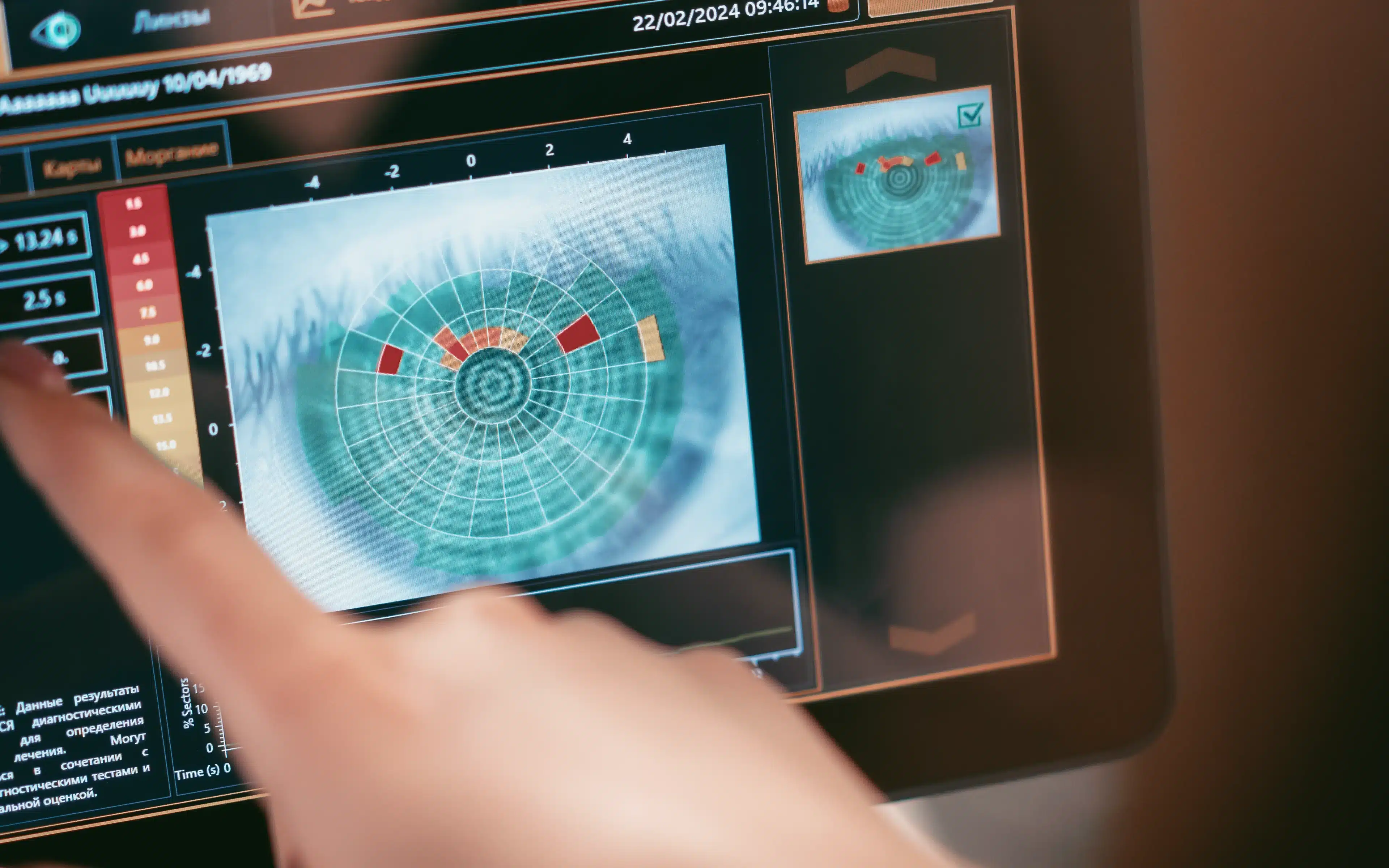
Tests to confirm juvenile retinoschisis begin with conventional eye tests followed by a field of vision test. Detailed assessment of the retina can be carried out using an ERG – an electroretinogram – that accurately measures reactions to light stimulus.
Treatment
There is no effective treatment of juvenile retinoschisis, even though attempts are made using laser surgery techniques. A programme of follow ups after initial diagnosis (at least every six months) is usually specified by the consulting ophthalmologist to keep track of any further degeneration.
In the event of severe loss of vision surgery may be undertaken to prevent an occurrence of retinal detachment.
Future developments
Research is currently underway via survey and blood samples to establish genetic functioning, examining young male sufferers but also female carriers.
With all the progress being made today in understanding the behaviour of genes in relation to how everything in the body works, it is to be hoped that gene therapy can be used in the future to provide effective treatment, especially considering the well known genetic path of the disorder.