
Words
Published
Retinal Detachment is an eye disease in which all or part of the nerve-fibre layer becomes detached from the back of the eye. As this layer is responsible for communicating visual images to the brain, this can be very serious.
It can also lead to the blurring of your vision and even a complete loss of eyesight. Retinal detachment may occur when the vitreous humor, the jelly-like substance responsible for maintaining the shape of the eyeball and for filling the space between the lens and the retina, gets pulled away and tears the retina in one or more places. While the pulling away of the vitreous humor is normal as a person ages, there are times when it gets stretched too much, thereby leading to the eye disease as fluid seeps through the tear to lift and detach the retina off the back of the eye.
Before the actual onset of retinal detachment, you will probably experience the posterior detachment of your vitreous humor first. When this happens, you will feel heaviness in your eyes. Light flashes will also bring about eye floaters.
This eye disease usually afflicts the middle-aged and elderly. Those with severe forms of myopia and those who have undergone cataract surgery are also vulnerable. Eye trauma resulting from a hard hit, like in boxing or other martial arts, may also lead to retinal detachment.
Symptoms of Retinal Detachment
If you are suffering from this eye disease, you will probably experience the following:
1. A loss of your central vision
2. A shadow in your peripheral vision that will slowly move to the center
3. Straight markers will look like curved lines
4. Extremely blurred vision, as if a curtain is covering it
Diagnosing Retinal Detachment
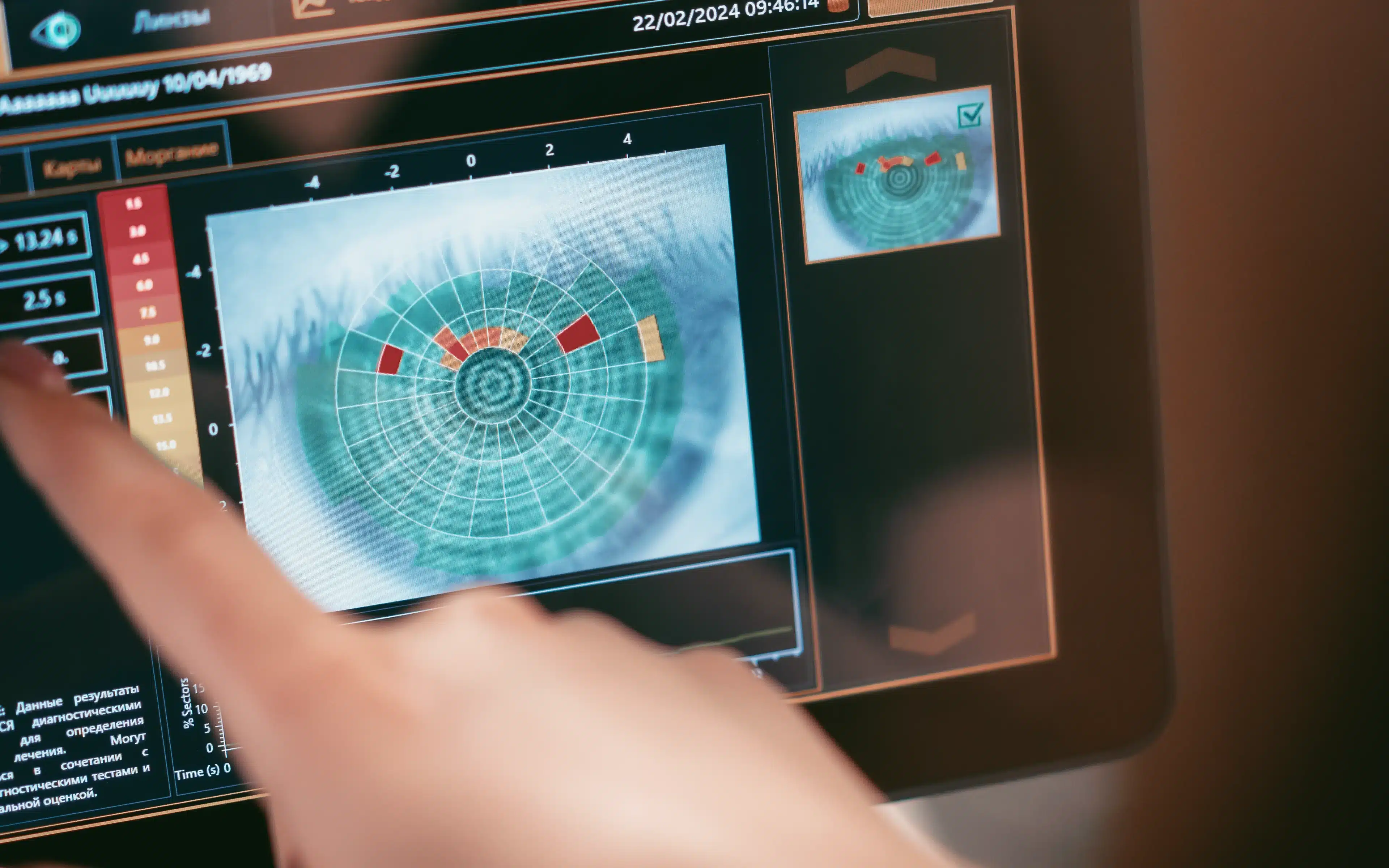
This eye disease may be diagnosed by your optometrist using a variety of tests. Among these are:
1. Flourescein angiography – this test involves photographing your retina by using a dye.
2. Ophthalmoscopy
3. Slit Lamp Examination
4. Ultrasound
Your optometrist may perform other tests in conjunction with the ones listed above to ensure a correct and proper diagnosis of this eye disease. Among the possible additional tests are:
1. Color perception Test
2. Electroretinography Test
3. Pupillary Reflex Response
4. Refraction Test
Treatment of Retinal Detachment
You should avoid activities that may increase pressure to your eye to lessen the chance of contracting this eye disease. This includes bungee jumping, diving, rollercoaster rides and skydiving.
If you do get this disease, your eye doctor will offer the following treatment options:
1. Cryotherapy, or freezing
2. Laser surgery. Expect to feel some discomfort after the procedure. Medications may be prescribed to relieve the pain and promote faster healing by your eye doctor, and a patch may be required to protect the affected eye.
3. Pneumatic retinopexy, in which a gas bubble is injected into the vitreous space of your eye
4. Putting a flexible band around the eye’s equator
5. Vitrectomy, or the removal of the vitreous gel that is pulling on your retina
If you are at risk of retinal detachment make an appointment with Dr Jim Kokkinakis at The Eye Practice for a comprehensive risk assessment on (02) 9290 1899 or book an appointment on line.


I had retinal detachment in another country but want to have a follow-up examination in Australia