Words
Published
Looking for the most comprehensive eye examination in Australia? At The Eye Practice, we’re committed to staying at the forefront of technology for diagnosing and treating eye disease. Our Sydney CBD practice invests in the world’s best equipment, providing comprehensive eye examinations using cutting-edge technology to assess and protect your vision.
Specialised dry eye assessment
Dr. Jim Kokkinakis has developed particular expertise in dry eye treatment, with patients travelling from Perth, Melbourne, and even New Zealand to our Sydney Dry Eye Clinic. What sets us apart is the advanced technology we use to diagnose and treat this increasingly common condition, which affects millions of Australians and is worsened by our harsh climate, air conditioning, and prolonged screen use.
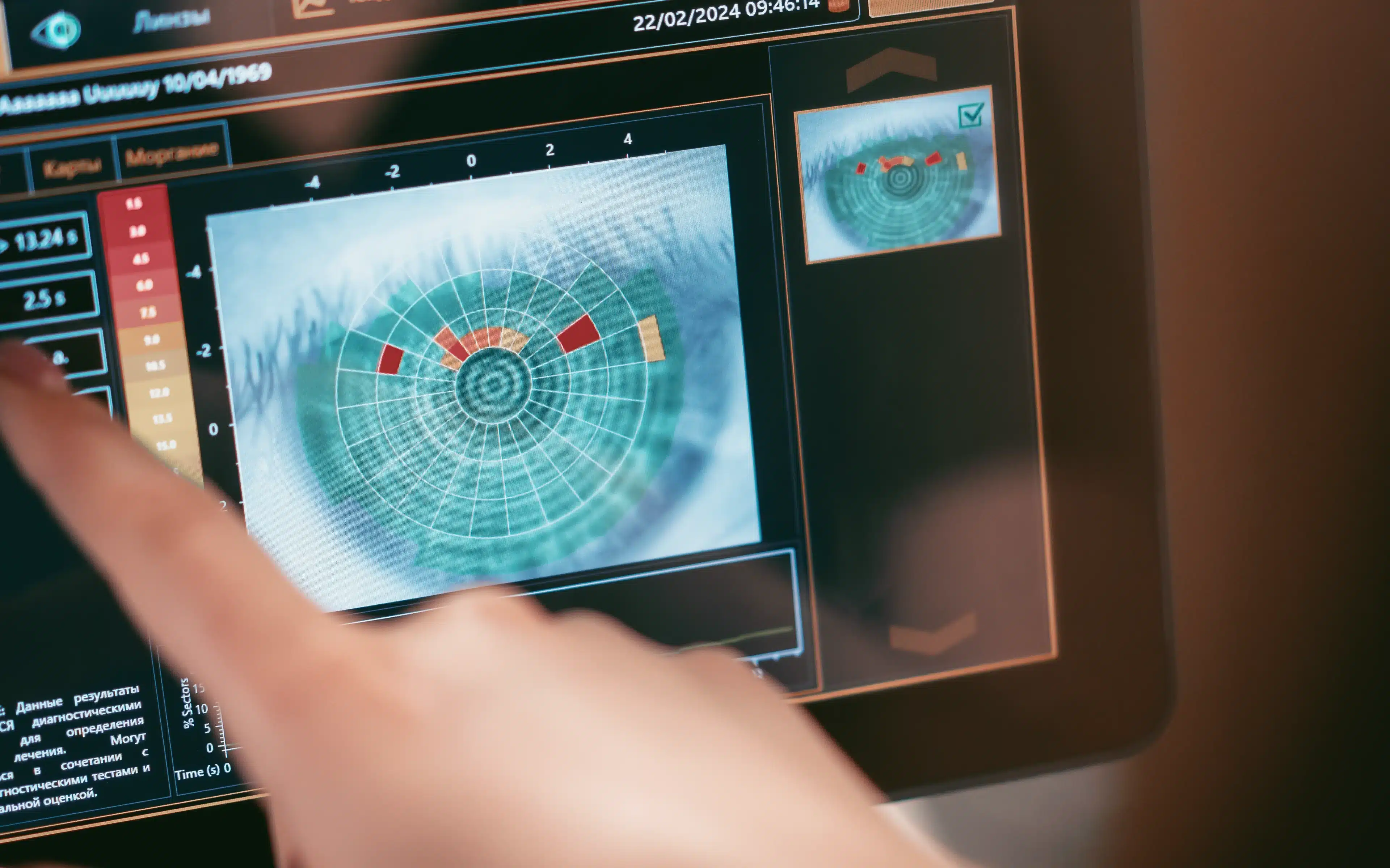
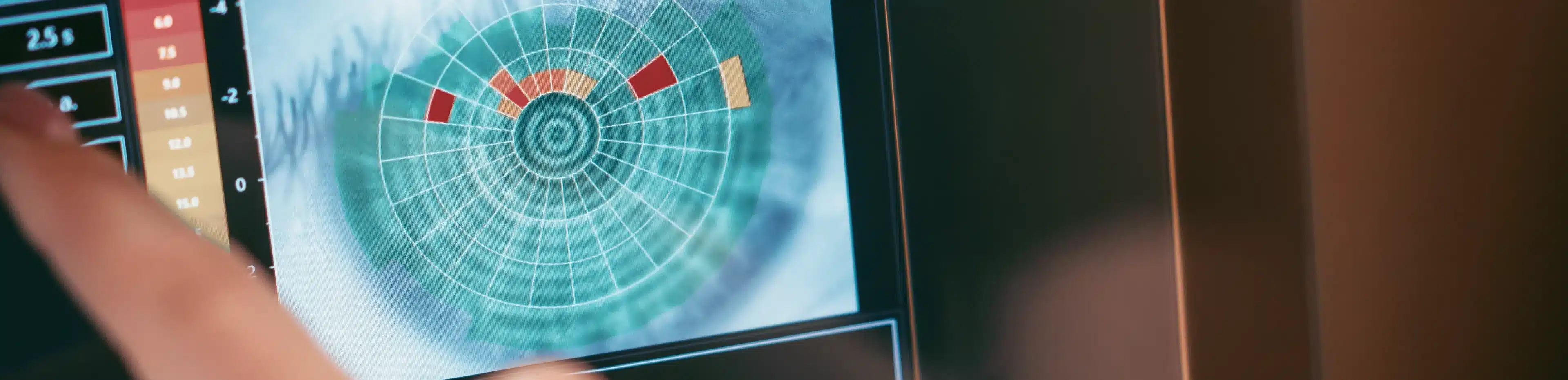
Our LipiView system measures two critical factors. First, it captures a 19-second video analysing your blinking frequency and completeness – essential for creating and distributing the tear film that lubricates and protects your eyes from infection and irritation. Infrequent or incomplete blinking is a major cause of dry eye and ocular surface disease. Second, it measures your lipid layer thickness, an important component of the tear film. Over 80% of dry eye problems relate to an unstable lipid layer, which allows excessive evaporation. We can monitor this thickness over time to assess how well our treatments are working.
For treatment, we use LipiFlow technology, which addresses meibomian gland dysfunction by simultaneously heating and massaging the meibomian glands. There’s no other technology like this, and we’re proud to be one of only a few practices in Australia offering this advanced treatment.
Capturing your retinal health
We routinely provide digital retinal imaging for all patients. Our digital retinal camera captures high-resolution images of the back of your eye, allowing a more detailed and wider-angled view than conventional observation methods. Even the most subtle, early signs of an eye condition can be detected.
These photographs provide a baseline for your eye health, making future changes much easier to monitor. This technology proves especially valuable in detecting and monitoring slow-developing, sight-threatening conditions such as glaucoma and macular degeneration, plus the eye-related effects of high blood pressure, high cholesterol, and diabetes. Your retinal images remain on file for direct comparisons from visit to visit.
Optical coherence tomography – seeing beneath the surface
Our OCT system takes comprehensive eye examinations to another level, detecting early signs of eye conditions before they affect your eyesight. This diagnostic imaging device captures cross-sectional images of the retina – the inner back surface of your eye. Our highly trained optometrists can detect early signs of retinal disease, glaucoma, and macular degeneration.
We also perform OCT on the front segment of your eye, including the cornea, iris, and lens. This high-resolution imaging system accurately measures corneal thickness before and after laser vision correction. It provides the most detailed cross-sectional view of the cornea, proving highly useful in monitoring conditions like keratoconus.
Precision contact lens fitting with corneal topography
As a practice heavily involved with contact lenses, we use corneal topography to map the shape of your cornea. This technology provides our optometrists with a three-dimensional representation of your eye, allowing us to choose the most suitable contact lenses from stock or have them custom-made at the laboratory. This ensures your contact lenses fit comfortably and provide the clearest vision possible.
Corneal topography also proves essential for monitoring degenerative diseases such as keratoconus and managing patients who’ve had corneal transplants. Australia’s intense UV exposure makes monitoring corneal health particularly important.
Visual field assessment – protecting your peripheral vision
In the early stages of glaucoma, most people remain unaware they’re developing the condition. Our detailed visual field assessment enables early detection of visual field defects, improving treatment success in preventing vision loss.
We perform visual field assessments on virtually every patient. This reflects our belief that an eye examination isn’t just about reading a letter chart, which only accounts for the central 2% of your vision. The other 98% of your vision – your peripheral vision – is equally important for daily activities, driving, and overall quality of life.