
Words
Published
There is still no ‘cure’ for keratoconus in the strict sense of the word. But latest high tech treatments for this disease are becoming more successful every year.
And there’s plenty more good news…
Contact lenses have never been more comfortable or provided better vision. In fact, this may be one reason why fewer people need a corneal graft every year now. New therapies promise a bright future for young people diagnosed today with keratoconus.
This post takes an in-depth look at seven of the latest bleeding-edge technologies for managing this eye condition:
The gold standard: corneal cross linking
Scleral mapping (or the 96-slice pizza)
I can see clearly now: dealing with optical aberrations
Eye drops to strengthen the cornea. human trials next
Ditch the itch: the latest anti-allergy treatment
Topography and the art of corneal mapping
1. The Gold standard: corneal collagen cross linking
There’s nothing new or controversial about corneal collagen cross linking. This procedure has revolutionised the management of keratoconus over the past decade.
Collagen strands make up the bulk of the cornea. They are like pipes stacked in a builder’s yard – orderly but free to move. As we age, the collagen strands form bonds with nearby strands. This is called cross-linking and is a natural part of the maturing of the eye.
The collagen cross linking procedure combines Riboflavin (Vitamin B2) and UV light to prematurely cross-link the cornea. This usually prevents (or greatly reduces) any further progression of the disease.
What does this mean?
One of the consequences of the widespread uptake of this procedure is that fewer patients need a corneal transplant than ever before. (This is not the only reason: the availability of better contact lens designs has also reduced the number of grafts needed each year).
Protocols vary from one surgeon to the next. Traditional techniques involve first removing the top layer of the cornea (the epithelium) and then applying the Riboflavin. Newer techniques involve leaving the corneal surface intact.
If your disease is actively progressing (getting worse) then this is the time to have cross-linking. It is not something to put off for a few years. As keratoconus is usually triggered by puberty, many people undergoing cross-linking are still in their teens.
Having this procedure can stop the disease in its tracks. If you’ve been stable for a few years or are in your thirties or older, it’s far less likely that cross linking would be indicated for your eyes. This procedure doesn’t cure or fix anything; it works by preventing further deterioration.
Vision can be hazy or blurred after cross linking for several months. It’s important to go into this procedure with reasonable expectations. You won’t get an improvement in your vision – in fact it may get a little worse before stabilising. But you will nip the disease in the bud and prevent further damage.
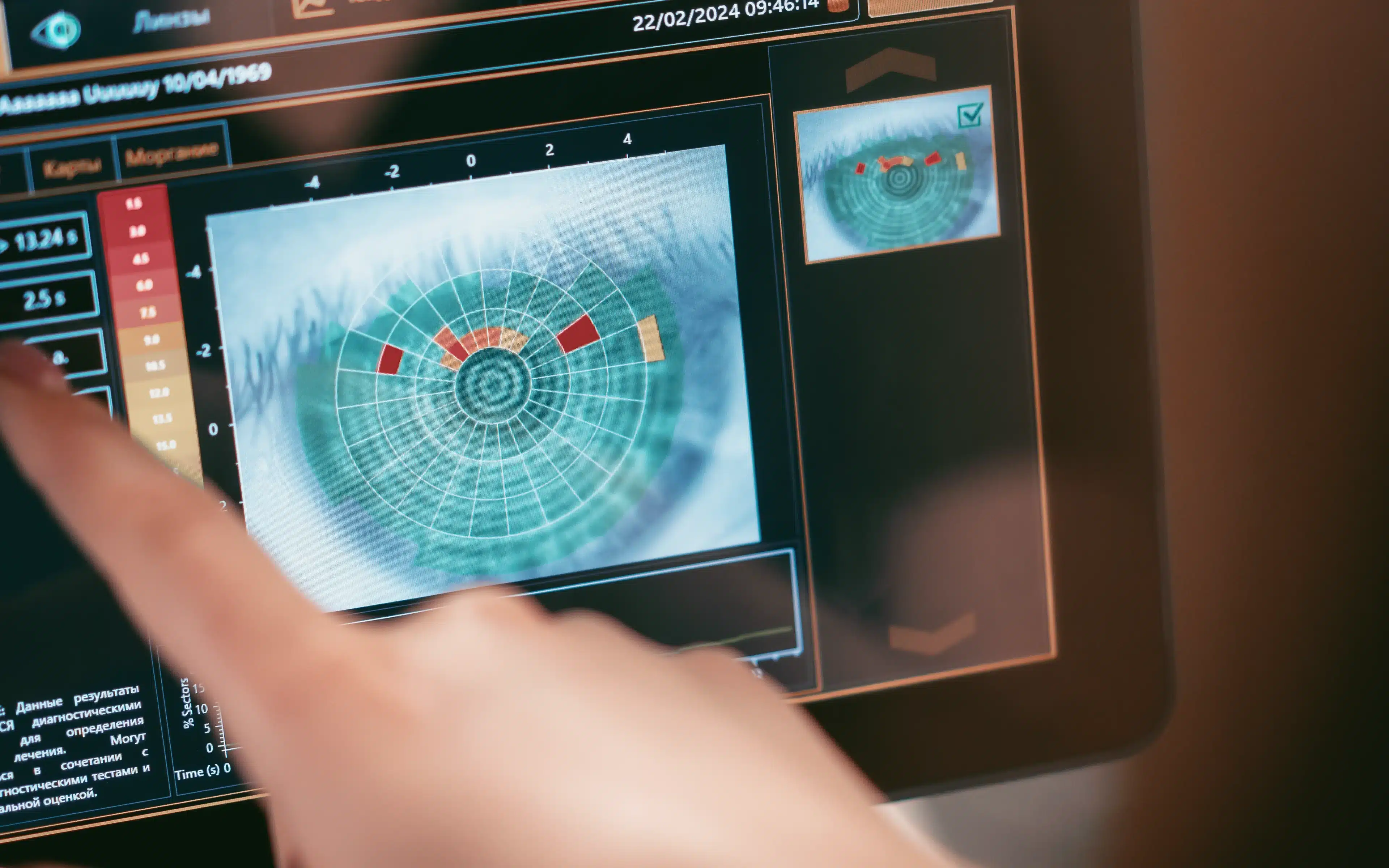
2. Scleral mapping (or the 96-slice pizza…)
Most people are familiar with the cornea – the clear front surface of the eye. The sclera is the rest of the eye’s surface – the white of the eye.
The sclera consists of a tough rubbery layer that protects the delicate structures of the eye. Scleral contact lenses are large enough to completely vault the sensitive cornea without touching it. Instead, they bear their weight on the white (sclera).
When properly fitted, these lenses are very comfortable and provide excellent vision. They keep many patients from needing a corneal transplant. In fact, the rate of corneal transplant has halved in recent years due in part to the uptake of scleral lenses.
Fits like a glove: the new scleral contact lenses
In the past, scleral lenses were notoriously difficult to fit. But in the last 12 months, scleral mapping has greatly improved this by allowing us to take better measurements of the eye’s surface.
The latest scleral mapping technology takes measurements in 96 meridians instead of the previous two. If that sounds confusing, picture a pizza cut into quarters, where the four quarters are different flavours. This is what we used to have (up to only a year ago).
With modern scleral mapping we now have 96 different flavoured slices of pizza, allowing an endless variety of shapes. The inside surface of your scleral contact lens is tailored for an exact fit with your cornea. This is great news for contact lens experts and patients alike.
3. I can see clearly now: dealing with optical aberrations
As mentioned above, the inside surface of a scleral contact lens can be tailor-made to fit your unique eye. Up until now, the outside surface of theses lenses has been a regular shape. This assumes that the lens will sit on your eye perfectly centred and aligned.
The reality is that contact lenses swivel, tilt and decentre. This has a big impact on the optics of the lens, and, consequently, your vision. You can’t stop a lens from moving but if it does so in a consistent way on your particular eye, we can then compensate for those movements.
An aberrometer can measure these distortions and create an optical fingerprint unique to your individual cornea. The technology, which is also used in astronomy lenses, compensates for the position of the lens on your eye and allows crisp vision.
This technology is very new and hasn’t made it into the individual practice yet, but it is not far off.
4. Eye drops to strengthen the cornea: human trials next
A New Zealand team of researchers has developed a tissue-engineering approach to treating keratoconus.
Tissue engineering is the science of improving or replacing biological tissue using various growth factors.
In this case, eye drops containing low levels of steroid and growth factors are applied to the eye. This causes the cornea to thicken and stiffen without affecting the optical properties of the eye.
Having proven the concept in the lab, live animal studies are now underway. Human trials are in the pipeline.
The active ingredients in these eye drops have previously been used in human eyes individually but not in combination. This fact should help get this treatment approved for human trails sooner rather than later.
Watch this space!
5. Ditch the itch! The latest anti-allergy treatment
It is now well established that eye-rubbing is a significant cause of keratoconus progression. Put simply, this means that rubbing your eyes makes your keratoconus worse.
Cutting out the rubbing is essential to managing the disease. But people with this disease can find that very difficult!
Why is this?
Chances are, if you have this eye condition, you also have asthma, eczema, hayfever or other allergies. These are called ‘atopic’ conditions. They cause itching, inflammation and irritation. And this causes eye-rubbing.
Stopping the itch takes away the desperate need to rub your eyes and this is where the latest anti-allergy medications come in.
Itching is caused by a natural chemical called histamine. This is released from our white blood cells into our bloodstream and causes a number of effects including ITCH and watering.
Antihistamine drugs block histamine from causing itchy eyes. Mast cell stabilisers are another class of drug. Their action is to prevent the release of histamine from the blood cells in the first place.
The latest drugs combine the actions of these two families of drugs (antihistamines and mast cell stabilisers) to stop itch before it even starts. Your therapeutic optometrist may prescribe Patanol or Zaditen eye drops for use once or twice a day. This will help manage the itch and help you to stop rubbing your eyes.
Believe it or not, this is an essential step in getting your eye disease under control and avoiding a corneal graft.
6. Topography and the art of corneal mapping
Not that long ago, if you were developing keratoconus, chances were that it would not be picked up for years. Your glasses might have been changed frequently but it was hard for most optometrists to diagnose early disease.
Often the first indication of a problem would be when you went for a laser eye surgery consultation. One of the first tests they do is map the shape of your cornea. Looking at the map, it is immediately obvious if your cornea is normal or if it is thin and steep (as in keratoconus).
These days however, it is not unusual for the optometrist in the high street to have a topographer – an instrument to map the contours (or topography) of your cornea. Many practitioners routinely map their patient’s corneas at the first visit as a baseline.
This has led to an increase in the early diagnosis of this eye disease. It doesn’t mean more people are affected by keratoconus; it just means that we’re discovering it earlier.
If you have any family history of this eye condition or if you suffer from allergies or rub your eyes, make sure you go to an optometrist that routinely uses topography to pick up early changes to your cornea. The earlier this disease is diagnosed, the earlier it can be halted in its tracks with therapies such as collagen cross linking.
7. Gene therapy and keratoconus
There is strong evidence to suggest that genetics plays a role in the development of keratoconus. Unlike some diseases, where a single gene defect is the cause, keratoconus sufferers have a number of mutations over various genes. This makes it more complicated to treat.
The tissue of the cornea is, however, ideal for gene therapy and there have been some exciting advances in this space in the last year.
Gene therapy was first used successfully in 1990, and over the last 25 years thousands of clinical trials have been conducted. Gene therapy involves introducing DNA into a patient’s cells as a drug to treat disease.
Gene therapy is a way to fix genetic problems at their source. DNA is used to create a functional (normal) gene to replace a mutated (harmful) gene.
To do this it has to reach the damaged cells of the cornea, enter those cells and disrupt their activity. This is still an experimental technique but recent advances in the delivery of the DNA to the cells are very encouraging.
8. 3D corneal bio-printing
The very latest research published in May 2018 offers new hope for people with corneal diseases such as keratoconus or other corneal issues such as injury or scarring. A team of researchers in Newcastle (UK) have demonstrated that a human cornea can successfully be engineered using a 3D printer and bio-ink. The innovative bio-ink is made from a collagen gel containing corneal stem cells.
The patient’s eye is first scanned and mapped and this map is exported to the 3D printer. The bio-ink then builds up a new cornea layer by concentric layer. The stem cells thrive in the gel and are nurtured to grow into a functioning cornea.
This technology is very new but holds great promise for millions of people who are blind from corneal issues. Further trials need to occur over the next few years before this can become a reality for real patients. It is a very exciting development so watch this space.
The take home message…
If you’ve been diagnosed with keratoconus, the 3 keys to a successful outcome are:
- early diagnosis
- referral for cross linking (if the disease is progressing)
- a comfortable solution for your visual needs. This may be the right pair of glasses or one of a wide range of contact lens options.
Make sure you see an optometrist who is highly experienced in keratoconus and sees this disease every day, not just once or twice a year.
They will have the latest technology to diagnose and manage your disease and they will keep up to date about new therapies and contact lens designs.
It is also important to realise that the vast majority of people do not need a corneal graft and actually do very well in contact lenses fitted by an expert.


Please add me to your mailing list
Grant I had a look at our website and thank you for point out that subscribing to our mailing list was not user friendly at all.
If I now can direct you to:
https://www.theeyepractice.com.au/optometrist-sydney/
You will find a subscription button near the top of the page.
H, thank you for this useful information, my son is 18yrs and has just had corneal collagen cross-linking procedure done for Keratoconus, it has been nearly 2 weeks and he had a setback with a blister forming. If there is one thing that I say to anyone who has had this procedure done, ice packs were the most helpful pain relief for him, he is now starting to see a little and feeling much better, still have a contact in and drops etc.