
Optical coherence tomography – or OCT – is a medical imaging technique. It uses visible light to produce a 3-D image of what’s happening beneath the surface. MRI, CT-scans and X-rays use radiation for this. But this can pose health risks, especially if repeated.
OCT is more like ultrasound. It only uses visible light (rather than sound). As a result, it is safe to use as often as you like without risk to the eye’s delicate tissues. It also provides much higher resolution.
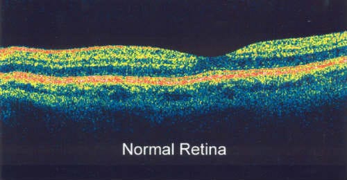
Some eye disorders are not visible with the naked eye. Indeed, the normal view or photograph that happens in eye examination does not show what’s happening under the surface. To see what’s really going on, we use OCT. It allows us to image the underlying layers of the eye in a non-invasive way. This is invaluable for picking up problems with the retina and macula as well as diagnosing and monitoring the progress of progressive diseases such as glaucoma.
How does OCT work?
OCT uses light from the red end of the spectrum (near infra-red). As a result, it can penetrate the light-scattering tissues of the eye (and other organs). Transparent tissues, like the cornea and lens of the eye, allow light to pass through. But retina and skin tissue are semi-translucent. This means only some light can penetrate. The long-wave light used in OCT bounces in different directions depending on what it comes up against. The scatter pattern builds up a picture of what lies beneath. (Ultrasound uses sound waves in a similar way).
Reasons for having OCT
Optometrists use optical coherence tomography (OCT) in the diagnosis and monitoring of glaucoma, as well as other eye diseases. It enables them to map the profile of the optic nerve. Information is stored so it can be compared to future scans to see if the disease is progressing or stable. Nothing gives a picture of what’s going on in your eye quite like OCT. That’s why The Eye Practice routinely performs this test as part of their comprehensive eye examination. We firmly believe that ‘an ounce of prevention is worth a pound of cure.’
Some eye conditions that are diagnosed or monitored using OCT:
- Retinal tear / detachment
- Macular hole / pucker
- Macular edema
- Age-related macular degeneration
- Central serous retinopathy
- Diabetic retinopathy
- Pre-retinal membrane
- Choroidal melanoma
- Vitreous detachment
- Glaucoma
- Optic Neuritis
More recently, OCT has been used in other areas of the body. These include dermatology and cardiology for diagnosing skin and heart diseases.
Want more technical information on OCT? Click here.


